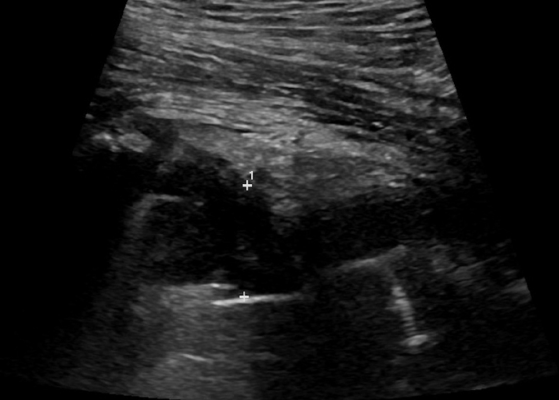
With hip ultrasound, you can see pictures of your hip muscles, tendons, ligaments, joints, bones, and soft tissues by using sound waves. The test may be used in infants to diagnose developmental dysplasia of the hip or to diagnose abnormalities. Noninvasive and ionizing radiation-free, ultrasound is a safe and noninvasive procedure.
There is little or no preparation required for this procedure. Wear loose, comfortable clothing and leave your jewelry at home. Dress in a gown if you are asked to do so. You will be instructed on how to prepare your child if they are being examined by a doctor.
You can find the best hip scan option at bothwellmedicalrooms.co.uk if you’re looking for a private scan.
Hip ultrasound imaging: what is it?
An ultrasound examination of the hip is known as hip ultrasound imaging. As an infant, the hips are mainly composed of cartilage and can be easily recognized on ultrasound scans since they have a ball and cup configuration.
The use of ultrasound imaging can be helpful to physicians in diagnosing and treating a variety of medical conditions, as it is non-invasive. In addition to being safe and painless, it is also quick. A sound wave creates a picture of what is inside the body. Also called sonography, ultrasound imaging uses sound waves to create images. Transducers and gel are directly applied to the skin using a small probe called a transducer. It is through the gel that high-frequency sound waves are transmitted from the probe into the body. Sounds that bounce back are collected by the probe. The sound waves are used by a computer to create images.
It is not necessary to use radiation during ultrasound exams (x-rays). Ultrasounds capture real-time images, allowing them to show how internal organs move and function. It is also possible to see blood flowing through blood vessels on these images.
Which preparations should I make?
You should wear comfortable clothing that fits loosely. If you are being examined in an area where clothing and jewelry may be necessary, please remove them.
The ultrasound examination process may be prolonged if the child is active or crying during the examination. Ultrasounds are highly sensitive to motion. A child should be explained the procedure prior to the exam to ensure a smooth experience. It would be helpful if you brought books, small toys, music, or games to keep the child occupied and make the time pass more quickly. A television may be present in the exam room. You can ask your child what his or her favorite channel is.
If your child is an infant, feeding him or her just before the exam may be helpful. Before feeding your baby, please check with the ultrasound staff to make sure that this is okay. Additionally, pacifiers or bottle feeding can be used during the examination. The infant may become more relaxed if you stand beside him or her during the procedure so he or she can see you and hear your voice.
What are the steps involved in the process?
In the same way that bats, ships, and fishermen use sonar, ultrasound imaging also works on the same principles. Sound waves bounce back or echo when they strike an object. The echo waves can be measured in order to determine the distance, shape, and consistency of the object. A solid object is one that is filled with fluid, while a fluid-filled object is one that is solid.
Ultrasound is used to detect abnormal masses, such as tumors, and to detect changes in organs, tissues, and vessels.
Transducers send sound waves and record the echo (returning) waves during ultrasound exams. Transducers send small, inaudible sound pulses into the body when pressed against the skin. Transducers record tiny changes in pitch and direction of sound waves as they bounce off internal organs, fluids, and tissues.
On a monitor, these signature waves are displayed as real-time pictures as soon as they are measured by a computer. In most cases, the technologist captures a frame or two of the moving picture as a still image. Also, the images may be saved as video loops.
What is the procedure?
In most hip ultrasound examinations, you will lie on your back or side on an examination table.
On the examination table, infants and children are usually lying on their backs for ultrasound studies.
During the exam, you will be positioned on a table by the radiologist (a physician who oversees and interprets radiological exams) or sonographer. To examine the body part under examination, a water-based gel will be applied. In order to make sure the transducer makes secure contact with the body, a gel will be applied. Additionally, it removes air pockets between the transducer and your skin that may block sound waves from entering your body. Once the transducer is placed on the body, it is moved back and forth over the area of interest until the images are captured.
The transducer is usually pressed against the area to be examined without causing any discomfort. The transducer can, however, cause minor discomfort if the area is tender.
A technologist will wipe your skin clean after the ultrasound imaging is complete. It won’t take long for any remaining portions to dry. There is usually no staining or discoloration of clothing caused by the ultrasound gel.
It is possible that the technologist will ask you to dress and wait while the images are reviewed once the ultrasound exam is complete.
How will the procedure affect me during and after it?
There is no pain or discomfort associated with most ultrasound exams.
Most ultrasound examinations are completed within 20 minutes, but it is possible for them to take longer sometimes. The initial review is sometimes followed by a few more pictures.
It is possible for the radiologist or sonographer to have you move the hip being examined during the hip ultrasound or may be able to move it for you to evaluate the functions of the joint, muscle, ligament, or tendon of the hip.
A hip examination is usually performed on both hips of an infant. There are a variety of positions and planes in which the hip is checked at rest and, occasionally, with gentle pressure. This is painless. The infant can be positioned with the help of a small towel.
A technologist will wipe your skin clean after the ultrasound imaging is complete. It won’t take long for any remaining portions to dry. There is usually no staining or discoloration of clothing caused by the ultrasound gel.
How does ultrasound imaging of the hip differ from other imaging techniques?
As ultrasound is not capable of penetrating bone, it can only visualize the bones on the outside, not what lies inside (except in infants whose bones are filled with cartilage). The internal structure of bones or certain joints is typically visualized by other imaging modalities, such as MRI.
For more news click thebritaintimes.co.uk